| dc.contributor.author | Demirel, A. Hakan | |
| dc.contributor.author | Kuşdemir, Ahmet | |
| dc.contributor.author | Kaptanoğlu, Buğra | |
| dc.contributor.author | Barlas, Mutlu | |
| dc.contributor.author | Bayram, Erkut | |
| dc.date | 2015-02-21 | |
| dc.date.accessioned | 2015-01-21T14:18:11Z | |
| dc.date.available | 2015-01-21T14:18:11Z | |
| dc.date.issued | 2003-01 | |
| dc.identifier.issn | 1302-4612 | |
| dc.identifier.uri | http://hdl.handle.net/11630/1640 | |
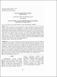
| dc.description.abstract | Anal kanala yerleşik nadir bir pilonidal sinüs antitesi tanımlanacaktır. Endoanal tip primer pilonidal sinüse oldukça nadir rastlanılmaktadır. 28 yaşında anal akıntı, kızarıklık şikayetleri ile başvuran erkek hasta 18 ay önce sakrokoksigeal pilonidal sinus tanısı alarak total eksizyon + Karydakis fleb ile tedavi edilmişti. Ameliyatta anal kanal muayenesinde kronik fissür gözlenmişti. Muayenesinde anal kanal posterior comissura’da 3 mm çapında dış ağzından pürülan hemorajik akıntı gelen, çok miktarda serbest kıl yapıları içeren sinus traktı saptandı. Metilen mavisi muayenesi ile ve kontrastlı grafide rektuma açılmadığı görüldü. Ameliyatta sinüs içeriği total olarak çıkarıldı, yara sekonder iyileşmeye bırakıldı. Histopatolojik tanı pilonidal sinus olarak belirlendi. Takdim edilen olgu endoanal tip pilonidal sinüs olup literatürde bildirilen 6.vakadır; etyolojisiııde kronik anal fissürün rol oynadığı görüşündeyiz. | en_US |
| dc.description.abstract | A rare case of a pilonidal sinus situated inside the anal canal is described. Endoanal type primary pilonidal sinus is rarely encountered. Twenty-eight years old male patient with the complaints of anal discharge and redness was diagnozed as sacrococcygeal pilonidal sinus and treated by total excision + Karydakis flep 18 month ago. By the operation during the anal canal examination chronic fissura was observed. During the examination we observed 3mm sized external orifice with a purulant hemorrhagic discharge at the posterior comissura of anal canal, the sinus tract was containing free hair bodies. When examined by methylen blue and contrasted x-ray graphy it is not found internal opening into the rectum. In the operation sinus was totally took out and wound was leaved seconder healing. Histopathologic diagnosis was pilonidal sinus. This is the endoanal type primary pilonidal sinus case and the sixth such case in the medical literature. | en_US |
| dc.language.iso | tur | en_US |
| dc.publisher | Afyon Kocatepe Üniversitesi, Kocatepe Tıp Dergisi | en_US |
| dc.rights | info:eu-repo/semantics/openAccess | en_US |
| dc.subject | Pilonidal Sinüs | en_US |
| dc.subject | Anal Kanal | en_US |
| dc.title | Anal kanal pilonidal sinüsü (Olgu Sunumu) | en_US |
| dc.title.alternative | A pilonidal sinüs of the anal canal (Case Report) | en_US |
| dc.type | article | en_US |
| dc.relation.journal | Afyon Kocatepe Üniversitesi, Kocatepe Tıp Dergisi | en_US |
| dc.department | Afyon Kocatepe Üniversitesi | en_US |
| dc.identifier.volume | 4 | en_US |
| dc.identifier.startpage | 63 | en_US |
| dc.identifier.endpage | 66 | en_US |
| dc.identifier.issue | 1 | en_US |
| dc.relation.publicationcategory | Makale - Ulusal Hakemli Dergi - Kurum Yayını | en_US |