| dc.contributor.author | Erkoç, Mustafa Fatih | |
| dc.contributor.author | Börekçi, Hasan | |
| dc.contributor.author | Sipahi, Mesut | |
| dc.contributor.author | Serin, Halil İbrahim | |
| dc.contributor.author | Akyüz, Yurdanur | |
| dc.date.accessioned | 2015-07-09T11:18:59Z | |
| dc.date.available | 2015-07-09T11:18:59Z | |
| dc.date.issued | 2015-04 | |
| dc.identifier.issn | 1302-4612 | |
| dc.identifier.uri | http://hdl.handle.net/11630/4095 | |
| dc.description.abstract | Amaç: Akut apandisit en sık karşılaşılan akut
karın tablosudur. Tanısı esas olarak öykü ve fizik
muayene bulguları ile konulmaktadır. Ancak
bazı vakalarda fizik muayene bulguları yetersiz
kalmakta ve arada kalınan vakalarda operasyon
kararı almak zorlaşmaktadır. Bu aşamada
bilgisayarlı tomografi (BT), ultrasonografi (US)
gibi radyolojik tetkikler ve laboratuar bulguları
tanıya yardımcı olmaktadır. Literatürde bu tanı
araçlarının birbirine üstünlüğü ve özgünlüğü
konusunda tam bir fikir birlikteliği yoktur. Bu
çalışmada amacımız akut apandisit tanısında
BT, US ve lökosit sayımının tanıya olan katkısını
karşılaştırmaktır.
Gereç-Yöntem: Çalışmaya son bir yıl içerisinde
AA tanısı ile opere edilmiş toplam 32 hasta retrospektif
olarak dâhil edildi. Hastaların US, BT
bulguları ve kan lökosit değerleri karşılaştırıldı.
Lökosit değeri 10.000/mm3’ün üzerinde olanlar
AA tanısı açısından pozitif kabul edildi.
Bulgular: 18 (%56.25) hastanın US bulguları, 28
hastanın (%87.5) BT bulgusu AA tanısı açısında
pozitif idi. 30 hastada (%93.75) kan lökosit de-
ğeri pozitif idi. Kan lökosit değerleri; US ve BT
bulguları ile karşılaştırıldığında istatistiksel olarak
anlamlıydı (p<0.05).
Sonuç: AA tanısında yüksek kan lökosit seviyelerinin
tanısal değeri hem US’ ye hem de BT’ ye
kıyasla daha yüksek bulunmuştur. | en_US |
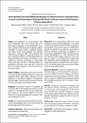
| dc.description.abstract | Objective: Acute appendicitis (AA) is the most
common cause of acute abdomen. It is diagnosed
with anamnesis and findings of physical
examination. But in some cases it is difficult to
decide for operation as physical examination
findings presents insufficient for diagnosis. In
such cases radiological studies including computed
tomography (CT) and ultrasound (US)
and blood sample analysis helps to correct diagnosis.
There is no consensus on superiority
and specificity of these diagnostic tools in literature.
We aimed to compare the contribution
of CT- US findings and leukocyte count on diagnosis
of acute appendicitis.
Material-Method: A total of 32 patients operated
with diagnosis of AA included to study respectively.
Patient’s US, CT findings and blood
leukocyte counts are compared. Patients with a
higher value of 10.000/mm3 leukocyte counts
are accepted as positive on the means of diagnosis
of AA.
Results: 18 patient’s (56.25%) US findings and
28 patient’s (87.5%) CT findings were positive
on the means of AA diagnosis. On the other
hand 30 (93.75%) patient’s leukocyte counts
were positive. Blood leukocyte counts were statistically
significant compared with US and CT
findings (p<0.05).
Conclusion: Diagnostic value of blood leukocyte
counts was found to be higher compared
to both US and CT findings in diagnosis of AA. | en_US |
| dc.language.iso | tur | en_US |
| dc.publisher | Afyon Kocatepe Üniversitesi, Kocatepe Tıp Dergisi | en_US |
| dc.rights | info:eu-repo/semantics/openAccess | en_US |
| dc.subject | Bilgisayarlı Tomografi | en_US |
| dc.subject | Ultrasonografi | en_US |
| dc.subject | Akut Apandisit | en_US |
| dc.title | Akut apandisit tanısında radyolojik bulgular ile lökosit sayımının karşılaştırılması | en_US |
| dc.title.alternative | Comparison of radiological findings with blood leukocyte count in the diagnosis of acute appendicitis | en_US |
| dc.type | article | en_US |
| dc.relation.journal | Afyon Kocatepe Üniversitesi, Kocatepe Tıp Dergisi | en_US |
| dc.department | Bozok Üniversitesi, Tıp Fakültesi | en_US |
| dc.department | Bozok Üniversitesi, Tıp Fakültesi | |
| dc.identifier.volume | 16 | en_US |
| dc.identifier.startpage | 136 | en_US |
| dc.identifier.endpage | 139 | en_US |
| dc.identifier.issue | 2 | en_US |
| dc.relation.publicationcategory | Makale - Ulusal Hakemli Dergi - Kurum Yayını | en_US |